Showing posts with label Gynecology. Show all posts

Wednesday, January 28, 2015

CIN: Decision Making Tool

Students get bored with the same pattern of learning. Students are tech savvy these days, and prefer methods that involve electronic gadgets. Some of them like decision making tools. With these points in mind, I used Microsoft Powerpoint to make an interactive presentation on cervical intraepithelial neoplasia (CIN). When one plays it in a Powerpoint Viewer, it keeps offering different options. One has to select that option on each screen that applies to his/her own patient. Then the tool offers another screen with more options. Finally one reaches a recommendation for that patient. I used to do it in Visual Basic. But Powerpoint is easier to work in, does not involve compilation, and creating a setup file, and does not require the user to setup the file on his/her computer. Most people have Powerpoint or equivalent program installed on their computers. So additional software is not required. The starting screen of the tool looks like this.

I showed it to my students today when I taught them CIN. I promised to make it available to them, so that they could use it. It will help them learn the topic. It might inspire some of them to do better than that piece of software. You can click on the image above to download it.

Tuesday, November 25, 2014

Reverse Trend in Hysterectomy for Cervical Cancer

For those who don't know, hysterectomy means surgical removal of the uterus. When the body of the uterus is removed and the lower part called cervix is left behind, it is called as subtotal hysterectomy. In general it is recommended that the cervix be not left behind, because a cancer may develop in it at a later date, and then the treatment of that cancer becomes difficult. Only in difficult situations does one leave he cervix behind, such as when the urinary bladder or rectum is densely adherent to the cervix.
That woman came from a place in north India. Some surgeon had removed her uterus.
"Why was a hysterectomy done on you?" I asked her.
"The doctor said it was the beginning of a cancer in the cervix of my uterus" she said, and showed me her case paper. "But he did not remove the cervix." I checked her papers. A subtotal hysterectomy had indeed been done for cervical intraepithelial cancer.
"But if it was done for the beginning of a cancer of the cervix, the cervix had to be removed" I said with amazement.
"Yes, doctor" she said tearfully, "but he did not remove it. It seems they do hysterectomy like this on all patients there."
I checked the doctor's prescription. His degree was MS in general surgery. I could not understand why a general surgeon would perform a hysterectomy these days. It was the job of a gynecologist.
"Doctor, save me" she said. "I have two small children."
"Don't worry" I said. "We will remove that cervix. It does not look like it is cancerous." It wasn't. I performed a vaginal removal of the cervix. It was a little difficult, because the abdominal structures were stuck to its top, and they had to be protected during its removal. Later on when I related this story to a friend, he said,
"If a young woman has cervical cancer and she desires to have more babies, they perform a radical removal of the cervix and keep the body of the uterus behind. This seems to be exactly opposite. The surgeon removed the body and left behind the cervix which he believed to have a cancer."
"Yes. This is a reverse trend in hysterectomy for cervical cancer. I hope he does not get any more patients like this."

Friday, September 26, 2014

Tactile Diagnosis of Septate Uterus

Congenital malformations of the uterus can be diagnosed by imaging techniques like ultrasonography, CT or MRI. Ultrasonography is noninvasive and economical. So it is often done as one of the many tests done for gynecological disorders. CT and MRI are used only if some anatomical abnormality is suspected and cannot be diagnosed accurately by using ultrasonography.. So uterine malformations may not be diagnosed when a woman presents with infertility and her clinical findings are normal. Such patients are subjected to hysteroscopy and laparoscopy to detect causes of infertility. Hysteroscopy detects uterine septa and bicornuate uteri coincidentally, the conditions not being responsible for the infertility. Laparoscopy detects bicurnuate uteri and may help suspect presence of a septate uterus.
We had one such patient. Hysteroscopy was done by a junior resident and a junior consultant. They could not visualize the uterine cavity very well owing to failure to achieve a good distension of the uterine cavity. Then they performed a laparoscopy on her. When I looked at the screen of the endoscopy monitor, I remarked,
"That looks like a septate uterus. The two parts of the uterus are seen separated by a groove running between them in the sagittal plane."
"....." they were not totally convinced. But one does not usually disagree with the boss, I suppose. It looked like the following model, except that the groove was a little less pronounced.


"I cannot put in a hysteroscope now. You did not have good distension, and hence you did not even connect the camera to the endoscope. So I may not get satisfactory distension too. Besides there will be some bleeding from endometrial trauma by the uterine manipulator that you are using. That will make the fluid turbid and obscure vision. Now we have to feel the inside of the fundus with a curette when you curette out the endometrium."
The junior consultant tried that after chromopertubation test was done and was found to be positive.
"The contour of the funds is smooth" he said. "There is no septum inside."
I was convinced there would be a septum in there. So I took over. I put the curette in the left cornual area, and slowly moved it along the inside of the fundus towards the right cornu. It dipped in the midline and then traveled up again to the right cornu. The path was as shown below.


"There is a septum in there" I said. "Watch the movement of the handle of the curette that is seen from the outside."
They watched intently. The handle did move as the tip moved inside the uterine cavity.
"Yes, there is a septum" they agreed.
"This is the tactile method of diagnosing a septate or bicornuate uterus" I said.

Sunday, March 16, 2014

Septic Focus

There was a patient in the gynecology ward. She had a gynecological condition, for which she was being treated on indoor basis. She also had intermittent fever, which was not due to any gynecological cause. Usual causes of fever had been ruled out.
"Look for septic foci" I said. :Make references to medical, ENT, and dental departments. It does not look like septicemia, but get her blood culture done, since the medical Registrar will ask for it anyway."
The next day we saw this woman again during our ward round.
"Sir, the medical and ENT Registrars have not found any septic foci in her" I was told. "The dental Registrar has noted the call but not seen her yet."
"But does she have any dental sepsis on gross?" I asked. "We can do the basic checking ourselves." I turned to the woman and asked her to open her mouth. She did so. I bent down and peeped inside. There were two smooth gum ridges but no teeth.
"Where are the teeth?" I asked.
"There are no teeth" she said calmly.
All faculty and Resident doctors burst out laughing.
"You could have saved the dental Registrar a visit to our ward by looking at her teeth. If she has no teeth, she cannot have dental caries and periodontal disease" I said. I knew she could have it, if she had a root broken inside the tooth sockets, but she would have had pain and swelling of the gums too.
"I knew she had not teeth. I wanted the dental Registrar to look for sepsis in the gums"  the Registrar said lamely.

Friday, March 14, 2014

Sinus Closure

Sometimes an abdominal incision does not heal and it gapes open. Usually there is some infection, which needs to be cleared by local dressing with/without systemic antibiotics. When healthy granulation tissue forms on the raw surface, the wound is sutured in one layer, using simple No.1 monofilament polyamide sutures.
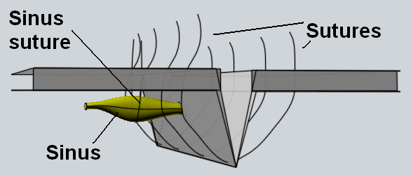
We had a case who developed MRSA infection. We controlled it with antibiotics. Healthy granulation tissue formed. But there was a problem. A tunnel of about 1-2 cm length had formed under the skin surface extending from the right side of the wound laterally in the subcutaneous tissue. It just would not fill up, and we could not pack it because its opening was not big enough. If we sutured just the original incision, this sinus would persist and keep discharging pus chronically. The option of laying it open by cutting healthy tissue overlying it was not very attractive, because that was healthy tissue and we did not want to disfigure the abdomen. Then I had an idea, which I have shown in the following 3D representation.
Four simple interrupted sutures of No.1 monofilament polyamide are shown. When tied, they will approximate the edges of the wound. The sinus is seen on one side, with a suture passing under it and exiting on either side of it over the skin surface. When tied, it will occlude the cavity of the sinus. Then it will heal because the granulation tissue within it will generate fibrous tissue to replace the cavity. The patient responded to this form of treatment very well.


Thursday, March 6, 2014

Tamponing

"Sir, we are tamponing this woman" I was informed during our round of the gynecological ward.
The word 'tamponing' reminded me of the word harpooning and then of captain Nemo and his Nautilus. Then it made me think of lampooning - which is transitive form of 'to lampoon', which means 'to attack or satirize in a lampoon', a lampoon being 'a satire in verse or prose ridiculing a person'. Then I thought of 'cartooning' meaning to draw a cartoon. But there is no such verb in the dictionary. I thought of all these other words because I believed there was no such verb as 'to tampon'. I checked the dictionary to make sure, and discovered that there was indeed a verb 'to tampon', which meant 'to plug a wound or body cavity with a tampon'. a tampon being 'a plug of lint, cotton or cotton wool, inserted into a wound or body cavity to stop the flow of blood or absorb blood. It is inserted by some women into the vagina during menstruation  to absorb menstrual blood. We use it in gynecology to reduce edema of the cervix due to pelvic organ prolapse. Of course it has to be soaked with glycerine-acriflavine prior to insertion for this indication.
This Resident Doctor made me proud by correctly using a word when all people are criticizing the Resident Doctors for using abbreviations which they make on the fly.

Thursday, February 20, 2014

Fluid for Distension of Foley's Balloon

Foley's catheter is a self retaining balloon catheter for constant drainage of the urinary bladder. There is a main channel, which drains the urine from the bladder. There is a side channel, which communicates with the balloon near the tip of the catheter. When it is in the bladder, it is inflated with sterile normal saline so that the catheter remains in the bladder. One uses sterile saline, so that if the balloon bursts accidentally, its contents will not contaminate the bladder and cause infection. Have a look at my 3D model of the catheter as shown below, before I tell you the story.
I was teaching the undergrads the technique of using a Foley's catheter. I taught them the theory, showed them a video, and then asked them to go see a catheter being passed in a patient preoperatively.. One of them had a difficulty.
"Sir, why use sterile normal saline?"
"What would you like to use instead?" I asked.
"Tap water" he said. "It is freely available, cheap and abundant."
"Won't the woman get cystitis if the balloon bursts and the unsterile water enters the bladder?" a girl from the same batch of students asked him before I could say anything. She seemed to be good at her studies and/or her thinking.
"That is right" I said. "Some people use any fluid anywhere. That can harm patients. I remember one of our professors using milk for checking tubal patency in place of methylene blue. Unfortunately the milk was not sterile evaporated milk, as is the recommendation. It was milk sold by the government's dairy. When I asked him about whether it could cause infection, he said, "No. It is sterile. It is pasteurized." It was sad that he did not know pasteurized milk was free of tubercle bacilli, but not of pathogens that can cause surgical infections. I don't know what happened to that patient, nor do I know what is happening to his patients where is practicing his art now."
They were suitably impressed.
"If we want to save some money by not using 5-10 ml of sterile normal saline" I said, "why not use the patient's urine? We could collect it after passing the catheter, and inject it into the balloon. Since it is the patient's own urine, it will not harm her even if the balloon bursts."
They laughed. No one gave the right answer to that question. Just then a senior consultant in the unit walked by, and said, "shall we put methylene blue solution into the woman's bladder? Then we will know immediately if the bladder gets opened."
I looked at the students, who caught on and laughed.
"Better not" I said. "A filled bladder is more likely to get injured than an empty one. Let us be more cautious during our surgery instead, and let us use methylene blue only if we suspect a bladder injury."
We completed the operation under discussion as usual without opening the bladder and there was no occasion to use any methylene blue solution or milk, and definitely not tap water.


Monday, February 10, 2014

Bonney's Hood Operation: Inverted T-shaped Incision

Bonney was a genius. His hood operation is an evidence of his genius. It is the only operation available for a posterior wall leiomyoma of the uterine corpus, that leaves the scar on the anterior wall of the uteurus, protected from adhesions with bowel and omentum postoperatively.
It sometimes so happens that the transverse incision on the corpus for enucleation of such a leiomyoma is not wide enough to get the leiomyoma out. Enlarging the incision laterally is not an option, because the uterus is only so much wide. We had such a patient the other day. The incision was not long enough to permit removal of the leiomyoma and there was no place laterally to extend it.. Then I had an idea.
"Make an inverted T-shaped incision" I said.
The operating surgeon, my Registrar made a vertical incision in the midline, starting upwards from the original uterine incision.
"Now raise the two triangular flaps upwards and outwards" I said.
That was done, and the leiomyoma came out. The flaps were too big for reconstruction of the uterus and had to be trimmed. The part cut in the midline got excised and we had a single flap to cover the surface of the uterus. I have shown the steps diagrammatically below, because there was no one to take pictures. Perhaps we will do that the next time.
Uterine incision
Vertical limb of the uterine incision
Flaps have been raised to expose the leiomyoma through an opening in serosa and pseudocapsule big enough to deliver the leiomyoma.


Saturday, February 8, 2014

Bonney's Myomectomy Clamp - Reverse Effect

Bonney's myomectomy clamp is used to temporarily occlude uterine blood vessels, so that there is little or no bleeding while the uterus is cut and leiomyomas within it are removed surgically. It is combined with application of an atraumatic clamp (sponge holding forceps) on each infundibulopelvic ligament to occlude the ovarian blood vessels which anastomose with the uterine vessels and supply blood to the uterus too.
We were performing a myomectomy for a large posterior fundal leiomyoma with encroachment on the upper part of the right broad ligament. Bonney's myomectomy clamp and sponge holding forceps were in place. When we opened the right broad ligament, even tiny blood vessels started bleeding furiously. Conventional methods proved inadequate to stop the bleeding. Then I had an idea. I released the Bonney's myomectomy clamp and sponge holding forceps. The bleeding stopped instantly.
"The clamp was occluding only the uterine veins, while the uterine arteries remained open and continued to pump blood into the uterus. That raised the venous pressure and caused such uncontrollable bleeding by back flow through the injured veins" I explained.
"We have not seen this before" they said.
"Nor have I" I said. "There is always a first time. Lucky for you it happened to someone else, so that you did not have to experience it first hand and try to find a solution to that problem."
We completed the myomectomy without any attempt at occluding uterine blood supply. There was not any hemorrhage.

Tuesday, January 28, 2014

Jinx of SUI

SUI is short for 'stress urinary incontinence'. It is a condition in which a woman loses a few drops of urine when she coughs, sneezes, laughs loudly or in severe cases, suddenly gets up from a lying down position. Victor Bonney described an ingenious test to diagnose it clinically, now popularly known as Bonney's test. In this test the woman in a dorsal examination position is asked to cough. If she loses a few drops of urine, the bladder neck area is elevated above the urogenital diaphragm by placing tips of index and middle fingers on either side of the urethrovesical junction. Then she is asked to cough again. If there is no loss of urine, the test is positive and the woman had SUI.
I have been performing Bonny's test for years. In the last few days something funny has been happening. There was a stout woman with symptoms suggestive of SUI. It is prudent to stand well away from the expected path of the urine that spurts out on coughing. I stood well away, and that too well on the right side rather than in front of her. When I asked her to cough, she coughed. The urine spurted out, but instead of remaining in the midline, it spurted to extreme right, exactly on my forearm, hand, and sleeve of my apron. I completed the test, washed away the urine and went to change my apron. A week later, there was another woman, of similar size and complaint. I was more cautious than the last time. I hid myself behind her leg and stayed as much to the right as was possible. When she coughed, her urine spurted more to the right than the last time. It soaked my forearm and hand, but spared my apron. I completed the test and washed away the urine. The third week, I decided that there was something wrong with the examination table and that caused a deviation of the stream of urine to the right. So I stood on the left side this time, well behind her left leg. When she coughed, the urine spurted to the left, on my left forearm, hand, and apron sleeve. I washed away the urine and changed my apron. But I could not get rid of a feeling that someone had put a jinx on that table. That jinx, which can be called the 'jinx of SUI', seemd to make the urine spurt on the examining hand, whether it was in midline, on the right or on the left. When I told my wife about this, she said with an amused look on her face,
"So what will you do now?"
"I have a plan to see what it exactly is" I said. "I plan to ask other clinicians if they have experienced this when examining patients on that table. If not, I will examine the next patient and then ask a colleague to examine her standing in the same position. If the urine spurts on both of us, the table is jinxed. If it spurts on my hand but not on my colleague's, someone has jinxed me."
"What if it spurts on your colleague's hand and not on yours?" she asked with the same amused look.
"Then one can say the jinx got confused or shifted from me to my colleague" I said.
She looked hard at my face and said nothing.
"Just kidding" I said.

Sunday, December 29, 2013

Round Ligament Confusion

The uterus has three structures attached to it near the cornu on each side, the round ligament in front, the fallopian tube in the middle, and the uteroovarian ligament behind. All three appear tubular (though only the fallopian tube is truly tubular, the other two are solid inside). One of them may be confused for any of the others, if the operative field is very small, as in tubal ligation during puerperium or during a minilaparotomy. Ligation of any of the two ligaments instead of the fallopian tube is a not infrequent cause of failure of female sterilization.
Usually this mistake is made by a junior resident doctor in training. He/she has to be reminded to trace the structure held laterally to see if it ends in fimbriae. If it does, it is truly the fallopian tube. During laparoscopic sterilization, it is often not possible to trace the tube laterally to see the fimbriae, because one performs it under local anesthesia (except in teaching hospitals) and insertion of multiple instruments is not possible. A rule of thumb is to visualize three tube-like structures at the cornu, the middle of which is the fallopian tube. If a person ranking higher than a resident doctor in training confuses something else for the fallopian tube, there is indeed good reason to get worried.
"Hey, look at the fallopian tube" exclaimed the Professor and Head of the unit while performing a laparoscopic sterilization. The fact that such a distinguished person found something different about a fallopian tube should suggest it was indeed different. I was a much junior person at that time. I put my eye to the eyepiece of the telescope.
"It is funny. It is curved and going to the internal inguinal ring" the Professor said.
The description was quite diagnostic and there was actually no need to look. It had to be the round ligament. I was already looking anyway and continued to do so as asked.
"There is another fallopian tube behind the one you have found" I declared politely after a respectable time interval. "That looks like a normal fallopian tube."
The Professor took charge of the laparoscope, looked inside, and ligated the tube without saying a word.
This must have been one of the many reasons the Professor considered me an enemy. I could have exclaimed "indeed it is an unusual fallopian tube" and allowed the Professor to ligate it. I would not have caused any feelings of enmity that way. I could not do it because I could not allow the poor woman to get pregnant due to ligation of a wrong structure.
I thought of this story after many years, when I saw another senior person (not as senior as that Professor though) make the same mistake the other day.

Sunday, December 22, 2013

Pouch of Douglas Occlusion

There was a patient who was undergoing an abdominal hysterectomy for menorrhagia and uterine leiomyomas. They called me half way through the surgery for an opinion.
"Sir, the rectum is adherent to the back of the supravaginal cervix. Shall we perform a subtotal hysterectomy and leave the rectum attached to the cervix undisturbed?"
I could understand the thought behind this suggestion. There was risk of rectal injury if one tried to separate it. Removal of the uterus above that level would get rid of the leiomyoma and the patient's problems."
I looked at the operative field. It looked like this (Sorry, there was no time to take a snapshot). I have drawn the structures as they would have been prior to any operative steps for better understanding of the readers.
The rectum was drawn up and stuck just above the anterior ends of both the uterosacral ligaments.
"Wait. Let me wash up and have a feel of it" I said.
I scrubbed and joined them. The rectum would indeed be injured if the uterosacral ligaments were clamped without separating it, and it could be injured during an attempt to separate it.
"I think I can get around this problem" I said.
Then I cut the posterior uterine wall transversely to a depth of 2-3 mm, just above the level of the uterine pedicles. I held the edges with Allis' forceps and cut sharply under the surface downward in the direction of the vagina. Once I reached the vagina, the posterior flap with the rectum attached to it could be pulled away from the uterus and uterosacral ligaments. I cut the ligaments within this cuff and ligated them. Then I cut the vagina all around and removed the uterus.
"This was somewhat like an intrafascial hysterectomy" I said. "The difference was that I kept a cuff of cervical fibrous tissue along with the fascial cuff to give additional protection to the rectum on the outside. After all, the serosa and fascia are thin and can get torn during dissection. Cervical fibrous tissue strengthened the cuff. Now will you close the vagina within this cuff? It won't injure the rectum, which is well away."
"Yes, Sir" they said happily.
I went away happy for having removed the patient's cervix successfully and also for having taught our doctors something new."

Sunday, December 1, 2013

Liquid Paraffin for Dressing: 2

I had written about how our Resident Doctors use liquid paraffin to dress an abdominal wound, when it is an adherent burst. For those of you who missed it, here is the link.
Liquid Paraffin for Dressing: 1
I had impressed on their minds that the commercially available preparation was not sterile and could not be used to dress wounds. I had told them to get it sterilized by putting it a hot air oven (dry heat) at 150 degrees Celsius for one hour. They did that for that patient.
There are two universal truths about things like this. One is that memories fade with time. The other is that history repeats itself.
"Sir, one patient with cesarean section has developed wound breakdown " the Registrar told me. "The surgeons have advised us to dress the wound with liquid paraffin."
"I trust you know how to sterilize liquid paraffin before using it for wound dressing" I said.
She looked at me blankly. One of the two universal truths had proved itself to be true.
"You have to put it a hot air oven (dry heat) at 150 degrees Celsius for one hour. Use it only after doing so" I said.
They looked at each other. After some time, they sent the Associate Professor to break it to me gently.
"Sir, they have ...um... already used liquid paraffin without sterilizing it. What shall we do now?"
They all kept looking at me, expecting me to explode. I have understood that anger does not do any good to anyone, and not desiring to be a victim of its effects myself, I spent time that I would take to count to ten.
"The germs have already passed into the wound" I said quietly. "See if you can put the whole woman in the hot air oven, which will sterilize the wound and the liquid paraffin in it."
They laughed. I thought it was sarcasm, while they thought it was a joke. The Associate Professor looked scandalized and worried.
"Don't worry" I told her. "I know they cannot follow my advice even if they try, because we do not have any oven big enough to take a human being."

Monday, November 11, 2013

FIGO Classification of Abnormal Uterine Bleeding

The International federation of Obstetrics and Gynecology has some pretty detailed stagings of gynecological cancers. They keep modifying these periodically. I had not thought they would go beyond that, but they did. I came across their classification of abnormal uterine bleeding (AUB) on the internet. Surprisingly it has not found its place in text books of Gynecology. I also found out that hardly anyone knew about it, though it came out in 2011. I tried to find out why. There were several reasons.
  1. They had done away with the age old terminology of menorrhagia, polymenorrhea, oligomenorrhea, metrorrhagia, hypomenorrhea, cryptomenorrhea etc, and replaced them with descriptive terms. The old terms were fine, brief, precise and well accepted. They caused no confusion and there was no need to change them.
  2. They had left things half way. While leiomyomas had been subclassified a lot, other lesions like polyps, adenomyosis had not been subclassified at all.
  3. They had pooled endometrial hyperplasia and malignancy together. Putting benign and malignant conditions of the uterus together made no sense.
  4. They had scrapped the term 'dysfunctional uterine bleeding' which was quite satisfactory.
  5. The purpose of this new classification was not clear. No purpose seems to have been served in the two years after they put out the classification.
  6. They put one condition - iatrogenic - in the list. They did not put any in which the patient ingested hormones on her own and caused abnormal uterine bleeding.
  7. They put a 'not yet classified' category. If they brainstormed and came up with a whole new classification which was to replace the old one, the new one had to be comprehensive. Leaving a big unclassified chunk does not make sense.
"Why have they done this?" someone asked me.
"To justify their existence?" someone else suggested. That sounded like justifying a trip to the city of the meeting, all expenses paid.
"To feel good at having created something new?" a third person suggested.
"To provide material for newer editions of textbooks?" someone snickered.
"I found the following explanation on the internet" I said. "These are quotes of the committee members.
  1. There has been general inconsistency in the nomenclature used to describe ...AUB in reproductive aged women, and there is a plethora of potential causes—several of which may coexist in a given individual.
  2. It seems clear that the development of consistent and universally accepted nomenclature is a step toward rectifying this unsatisfactory circumstance. Another requirement is the development of a classification system for the causes of AUB, which can be used by clinicians, investigators, and even patients themselves to facilitate communication, clinical care, and research.
  3. The goal of our panel was to develop an agreed pragmatic classification system with a standardized nomenclature to be used worldwide by researchers and clinicians investigating and treating women of reproductive age with AUB."
 They all were stunned.
"What does it all mean?" the first person asked.
"Does it mean anything?" the second person asked.
"It all reminds me of the preventive and social medicine text books. A lot of words, and you are blank after reading them" the third person said. I neither agreed nor disagreed with any comment.
"The concluding remarks included this line" I said. "We recommend a scheduled systematic review of the system on a regular basis by a permanent committee of an international organization such as FIGO."
"Now we know what it is all about" they said and laughed.

Saturday, November 9, 2013

SVP's Test

One of my old Resident Doctors was visiting us.
"We had fun" she said. "I still remember your SVP's test. Do you still do it?"
I smiled. I used to do things which were unorthodox, but would help the Residents remember things better. This test was to demonstrate how loose a ligature was, so that they would remember to tie tighter ligatures.
"No" I said. "The fun wears out if you do the same thing over and over."
After she went away, I sat thinking of the test. I used to call it SVP's test for want of a better term, not to immortalize my name. Anyway, there was nothing wrong with naming the test after me - after all I had developed it myself. Nothing great I suppose, but effective.
After a Resident Doctor had tied all ligatures while performing a hysterectomy, I would take a curved hemostat and try to pass it gently between the pedicle tied and the ligature. If it passed through, the ligature was loose. If it did not, the ligature was tight. Then I would make him/her put a figure-of-eight ligature over the ligated pedicle to avoid postoperative bleeding from the loosely ligated pedicle. Later I made it a policy to place figure-of-eight ligatures on all ligated pedicles for safety. Then this test became superfluous. I performed the test recently because a doctor trained in another institute wanted to do away with the placement of the safety ligatures - or rather did not know about them. The results are shown in the following picture.
The hemostat is seen to have passed through the ligatures on two pedicles. The test was thus positive on both the  pedicles tested. Needless to say, I made that doctor place safety ligatures on all pedicles and advised to do so in all cases operated on subsequently.

Friday, October 18, 2013

Lateral Myomectomy

I have developed a new concept for removal of uterine leiomyomas. I call it 'Lateral Myomectomy'. Conventionally abdominal myomectomy is performed through preferably anterior uterine wall incisions, and when that is not possible through posterior uterine wall incisions. In both of these cases, the uterine scar is exposed to the intraperitoneal structures and adhesions with bowel and/or omentum are quite likely. This risk can be reduced using barriers to cover the suture lines until the serosa heals, like hydrofloataion or oxidized cellulose cover. The best way would be to avoid a serosal incision, which I achieve by opening the broad ligament and cutting the lateral uterine wall.
This technique is applicable when there is a large leiomyoma that expands the uterus globally i.e. in all directions. That expands the lateral uterine wall too. If it is an anterior wall leiomyoma, the round ligament and anterior leaf of broad ligament are cut. The uterine incision is made anterior to the ascending uterine vessels, above the uterine artery stem. If it a posterior wall leiomyoma, the posterior leaf of broad ligament is opened in front of the attachment of the utero-ovarian ligament, and the uterine incision is made posterior to the ascending uterine vessels. In both the cases, the uterine vessels are protected as the incisions runs parallel to them. The ureter is protected because it lies below the uterine artery stem, and downward extension of the incision is avoided by application of an Allis' forceps at that end. Fallopian tube is protected by applying an Allis' forceps at the upper end of the incision to avoid an extension of the incision during removal of the leiomyoma.. Tunneling incisions can be made as required through the bed of the leiomyoma removed, so as to remove any other leiomyomas present. Closure is by conventional technique. At the end of the operation the divided round ligament is reapproximated, and broad ligament is closed. There is no scar on the serosa of the uterus.

I have put a video of this on YouTube for those who want to see it. Do write to me if you have any questions.
YouTube Link:  http://youtu.be/y1g5hPzHzw0

Monday, October 14, 2013

Free Intraperitoneal Cyst

A 45 years old woman presented with third degree uterine prolapse, cystocele, rectocele and lax perineum. She had had no symptoms of acute abdominal pain in the past. We performed a vaginal hysterectomy, anterior colporrhaphy, vault suspension and posterior colpoperineorrhaphy on her. After division of the uterine vessels, I noticed a dirty yellowish colored cystic structure above and behind the uterine corpus. I removed the uterus. Then I held the structure with Babcock's forceps and made traction. It delivered without any resistance. It had absolutely no attachment to any intraperitoneal structure.
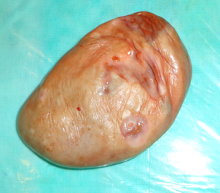
It was soft, cystic, flaccid, and tended to flatten out when placed on a flat surface. The surface did not show any place where a pedicle could have necrosed and set it free in the peritoneal cavity. I inspected both the ovaries and found them to be normal. I cannot put a picture of the operative field showing the ovaries because that would show body parts which may be considered objectionable by some viewers and also by Google who gives me free space for this blog. Histopathology of the cyst showed degenerated cyst possibly of ovarian origin.
"Sir, where did it come from?" a Resident doctor asked me.
"I would have said one of the ovaries. But the ovaries are all normal. An ovarian cyst develops within an ovary, not from the surface of an ovary with a pedicle that can undergo necrosis and set the cyst free. If the ovary with a cyst in it undergoes torsion and gets detached, the cyst may become free, but it would contain the ovary too, not leave it behind. It cannot have been a broad ligament cyst, because it was above and behind the uterus. If it had been in the broad ligament, we would have seen it before clamping the uterine vessels. Broad ligament cysts do not escape into the peritoneal cavity anyway."
"Why don't you publish it in a scientific journal?" a colleague asked me.
"These days many journals ask for a lot of money to publish articles. Those which do not do so ask silly questions about the content and their reviewers pass comments which if made by exam going students would result in them being failed. Anyway the readership of my blog is large enough for me to be happy as a teacher. I know how many people read my given article in my blog, which is not what I can say about my article in a local scientific journal. After all, a blog post disseminates scientific information as much as a scientific journal, if not more. It is free too, while most journals ask money from readers too."


Saturday, October 12, 2013

Migrating Leiomyomas

Birds migrate. People migrate. The human placenta migrates from lower uterine segment to upper uterine segment sometimes. But more about it some other time. This one is about migrating location of uterine leiomyomas.
That patient came to our outpatient clinic with about 9 cm diameter leiomyoma in the vagina. Since we could not reach the upper part of the leiomyoma, we could not see where the cervix was. She had an ultrasonographic report from an outside clinic showing that it was a central cervical leiomyoma arising from the anterior wall, stretching the posterior cervical wall over it. It did not disclose the location of the external os. Our people are intensely loyal to our institute. So they got a scan done by our sonographers. Their report came as a central cervical leiomyoma arising from the posterior wall, stretching the anterior cervical wall over it.
"Sir, the reports are exactly opposite of each other" my Registrar said.
"Could the origin of the leiomyoma have migrated between the two scans?" someone sniggered.
"I would tend to believe the first report, because it is done by a qualified sonologist, with some experience in private practice. The second report is by a resident doctor in training. We also have enough experience of our local people, who sometimes give mirror image locations of structures instead of their actual locations. We have to explore and remove the mass anyway, no matter where it is coming from. So relax. We will know about its location on our OT day."
It turned out to be neither. It was a polyp arising by a short pedicle from the posterior endocervix 0.5 cm above the external os. No part of the cervical canal was stretched over it."
"Our people were more right than the qualified private practitioner, though both were wrong" said someone. "How do we explain that?"
I was stumped.
"The only explanation is that he must have been one of our local people, now qualified and in private practice" someone said. Everybody laughed.
I knew it was not right to laugh. I also knew there was something that needed to be set right. But I could not do it because I was not a sonographer and did not know how.

Friday, October 4, 2013

Gloves Over Gloves

A large majority of doctors in my department wear two pairs of gloves during an operation. I had a general idea why they did so. But these are times of precision. So I asked people who worked in the OTs their reasons for doing so. I had to use intermediaries in case of residents for fear of not getting their true answers if I asked the question myself. Some of the answers were educative and entertaining at the same time. They were as follows.
  1. Hospital supply of gloves is such that one or both the gloves tear during use. When that happens, there is another one under it. That gives protection to ....(read answers 2 to 4).
  2. When the outer glove tears, the inner glove protects the doctor from catching some disease from the patient's body fluids.
  3. If I wear only one pair and one of the gloves tears, I get electric shock when I use electrocautery. If I have two pairs on, I don't get any shock when the outer glove tears, because the inner one electrically insulates my hand.
  4. When the outer glove tears, the inner glove prevents bacteria from the surgeon's hand entering the operative field.
  5. When the surgeon wears the gloves using a wrong technique, i.e. touching the outer surface of a glove with bare skin, it gets contaminated. Another glove on top of it nullifies the effect of this contamination.
  6. The air conditioner in the OT is so strong! Two gloves keep my hands warm (with the air cushion in between them as insulation?).
  7. After the operation, I remove the outer blood stained gloves and use the clean ones inside to apply dressing to the wound. That keeps the dressing clean.
  8. When I have to get an instrument from the general trolley while assisting an operation and there is no one to give it to me, I remove my outer glove, get the instrument, and then put on the glove again. Thus I do not contaminate the general trolley.
  9. I do it because the senior resident told me to do so.
  10. I heard the Boss hits on the knuckles with a surgical instrument if the resident goofs up while assisting him perform an operation. Two gloves would reduce the trauma. (Would knuckle pads be more useful?)
If you wear two pairs of gloves and have any reason other than those listed here, please write to me. I will update this post with the newer answers.

Tuesday, August 27, 2013

Medical Violence

The most common place of violence is in the movies. It is both general violence and violence against women. Going by media coverage and social media anger, the most common place for the violence against women would be Delhi and Mumbai, though by crime records it is anywhere in certain states in the country.
No one has talked about medical violence yet. That does not mean it does not exist. There is that occasional doctor who threatens to slap a woman screaming in pain in labor, unless she stops screaming. There was that House Officer a few years ago who had touched a woman's nose with the scissors he was holding while conducting childbirth, and said,
"Stop screaming or I will cut off your nose." She stopped, saved her nose, and now he is somewhere in UK. I trust he does not indulge in such practices there, or they would have deported him minus his nose long ago.
I have heard of House Officers who have slapped patients' thighs to stop non co-operation during childbirth.
"She was absolutely hysterical." the explanation would be given. "When I slapped her thigh (the only part nearby) she suddenly became co-operative and the delivery took place safely."
I had been aghast whenever I heard these stories. I thought nothing could be worse. I was wrong. Just the other day I heard another one that would beat any other story.
"He was Registrar when I was A House Officer" the story teller said. "He was quite rough on patients, many times abusive and at times violent. I remember two patients in particular. He had dislocated a patient's jaw once by slapping her face. The other one was given a broken finger. The reason was that they became hysterical with labor pains."
"Huh?" I was aghast again, this time more than at the previous times. "What does he do these days?" I thought he would have been stopped from practicing by some patient who went to a court against him.
"He is still around, practicing obstetrics and gynecology."
Lucky fellow and unlucky patients!

प्रशंसा करायचीय, नावे ठेवायचीयेत, काही विचारायचय, किंवा करायला आणखी चांगले काही सुचत नाहीये, तर क्लिक करा.

संपर्क